Pilonidal Cyst Pictures: Before, During & After Treatment
Real cleft lift before and after photos from Dr. Rafailov's Stanhope, NJ practice
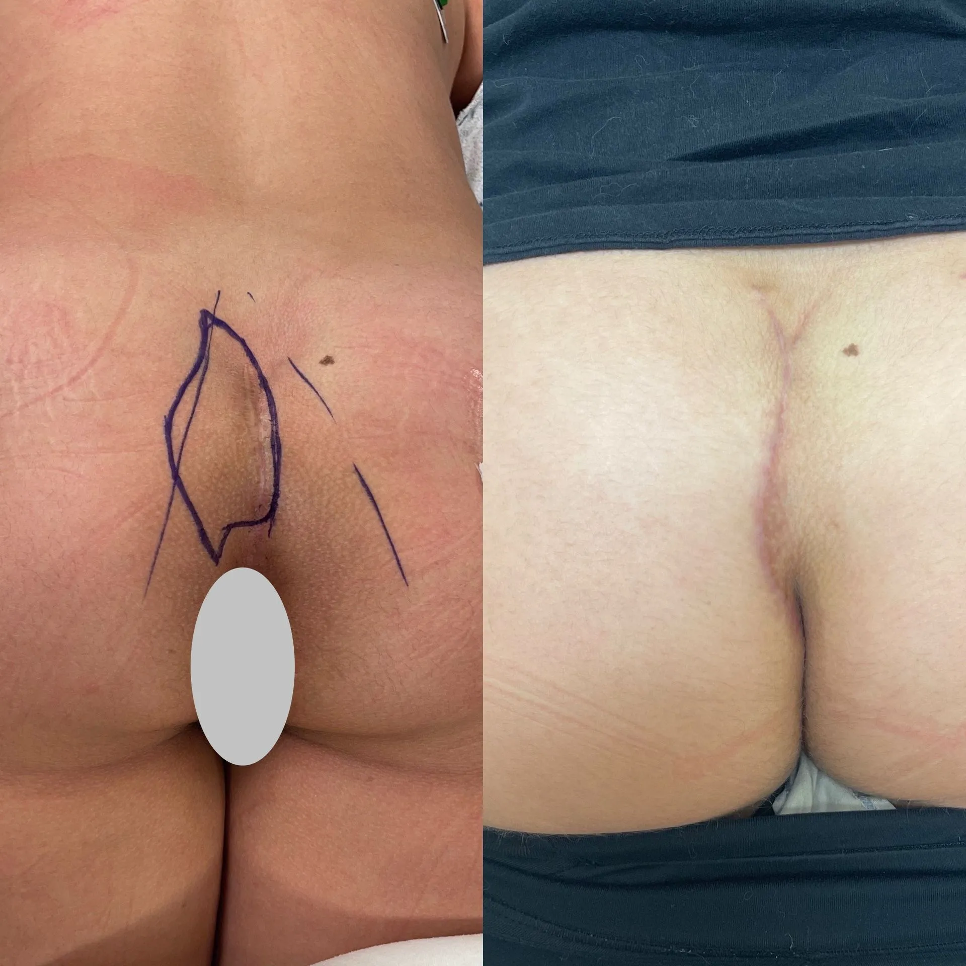
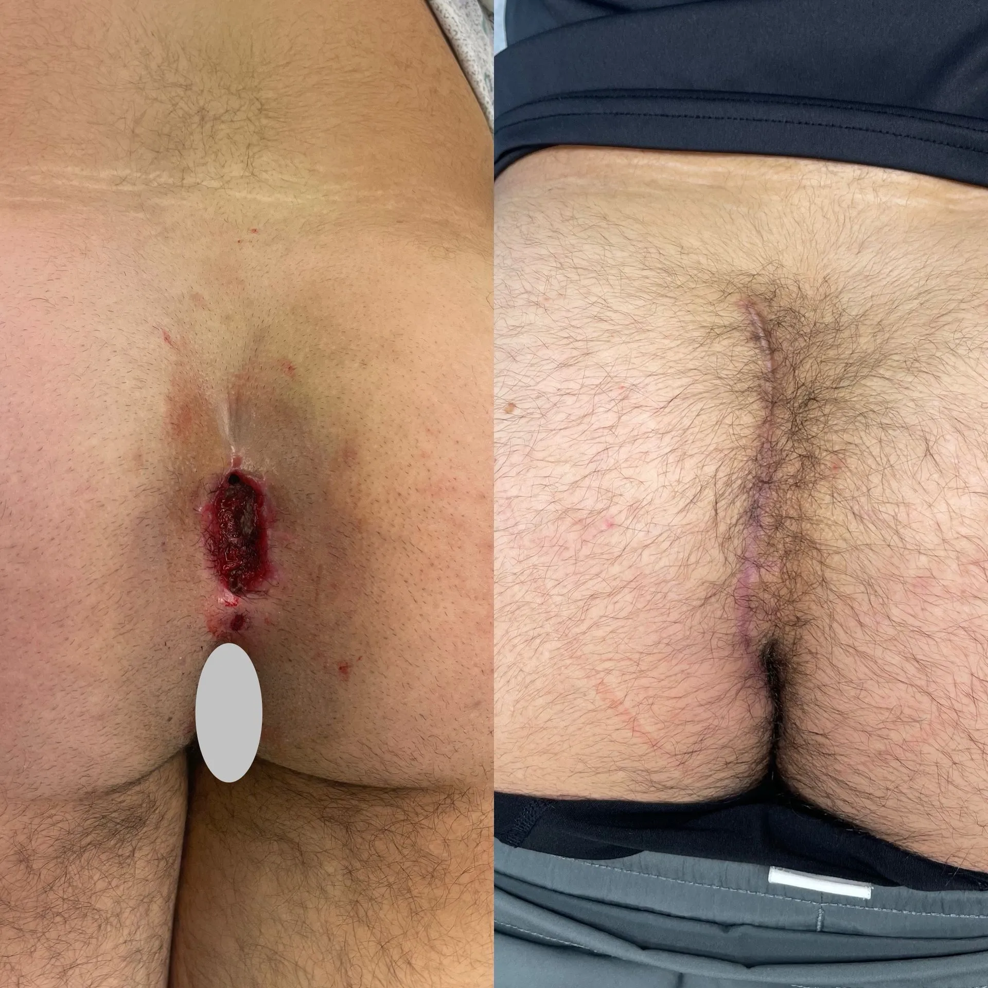
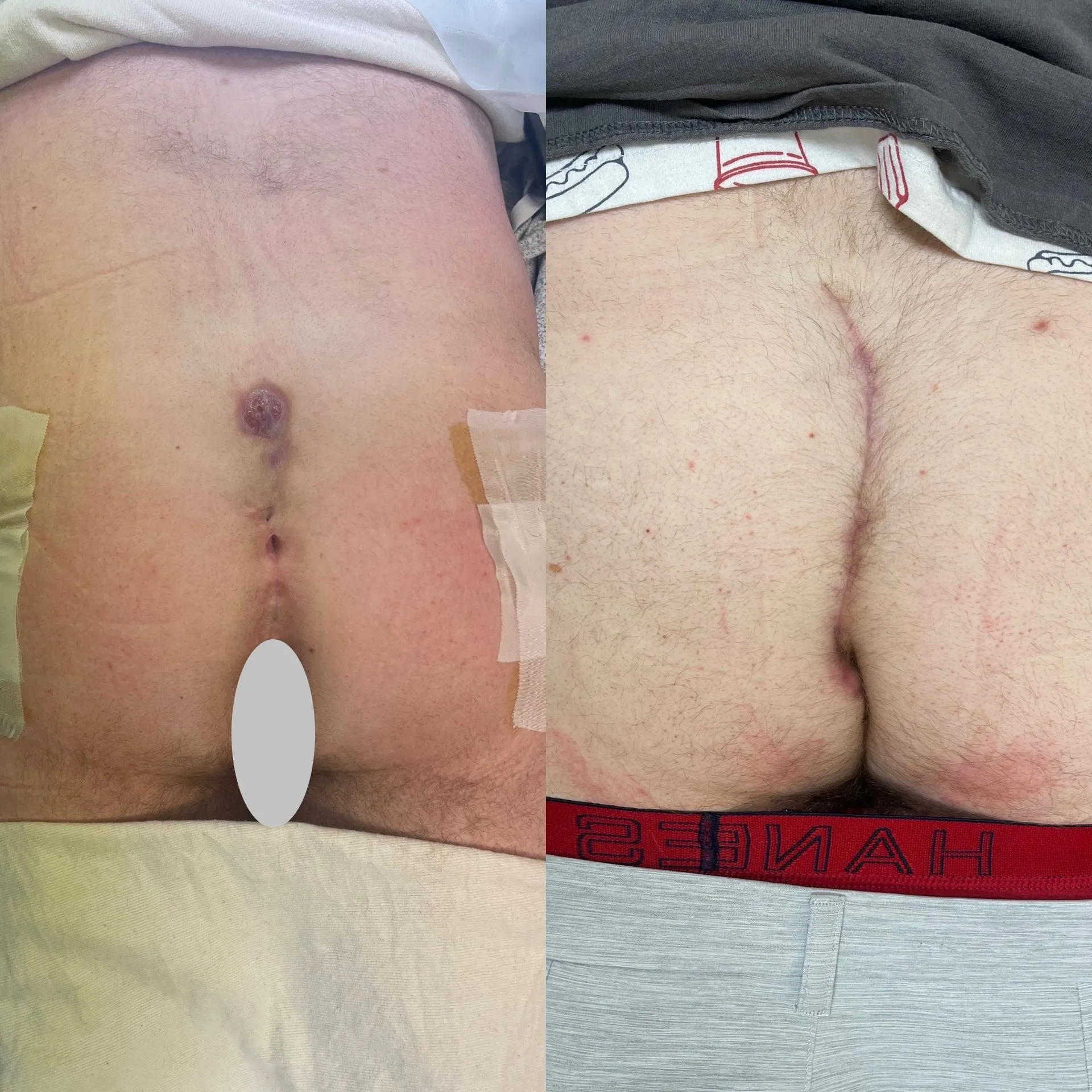
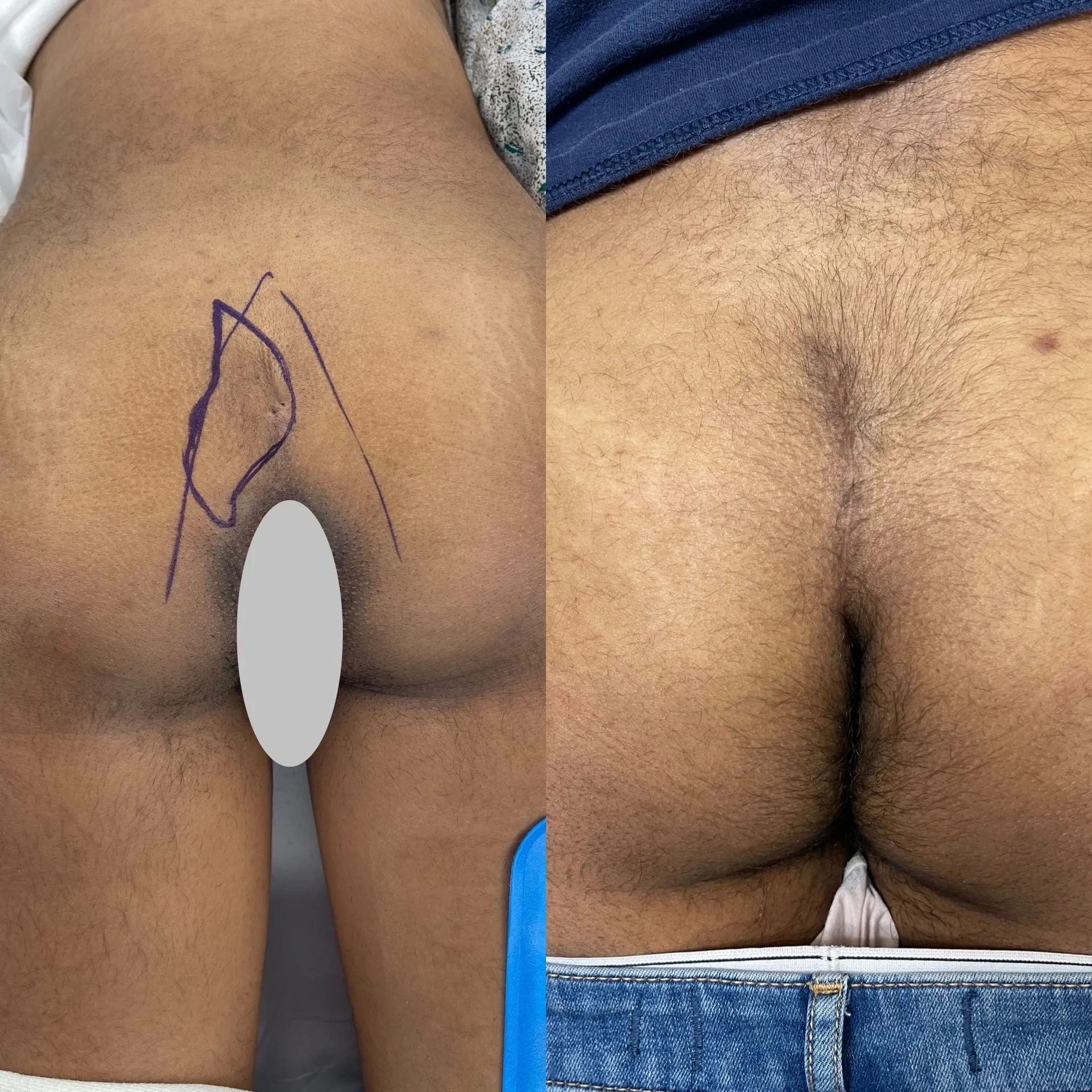
The photographs below document cleft lift surgery outcomes for patients treated by Dr. Samuil Rafailov for pilonidal disease. Each pair shows the affected area before surgery on the left and the same area after full healing on the right, typically photographed between 3 and 10 weeks post-op.
Pilonidal disease can present in several stages. Early-stage pilonidal cysts often appear as small midline pits with mild swelling or drainage. Abscessed or infected presentations involve a tender, red, fluid-filled mass that may rupture or require drainage. Recurrent disease shows scarring and repeat sinus tracts after prior procedures. The "before" images in this gallery capture a range of these presentations as they appeared at the time of consultation.
The "after" images focus on post-surgical healing following cleft lift reconstruction, the procedure Dr. Rafailov specializes in for definitive treatment. Viewer discretion is advised; these are clinical photographs of real patient results.
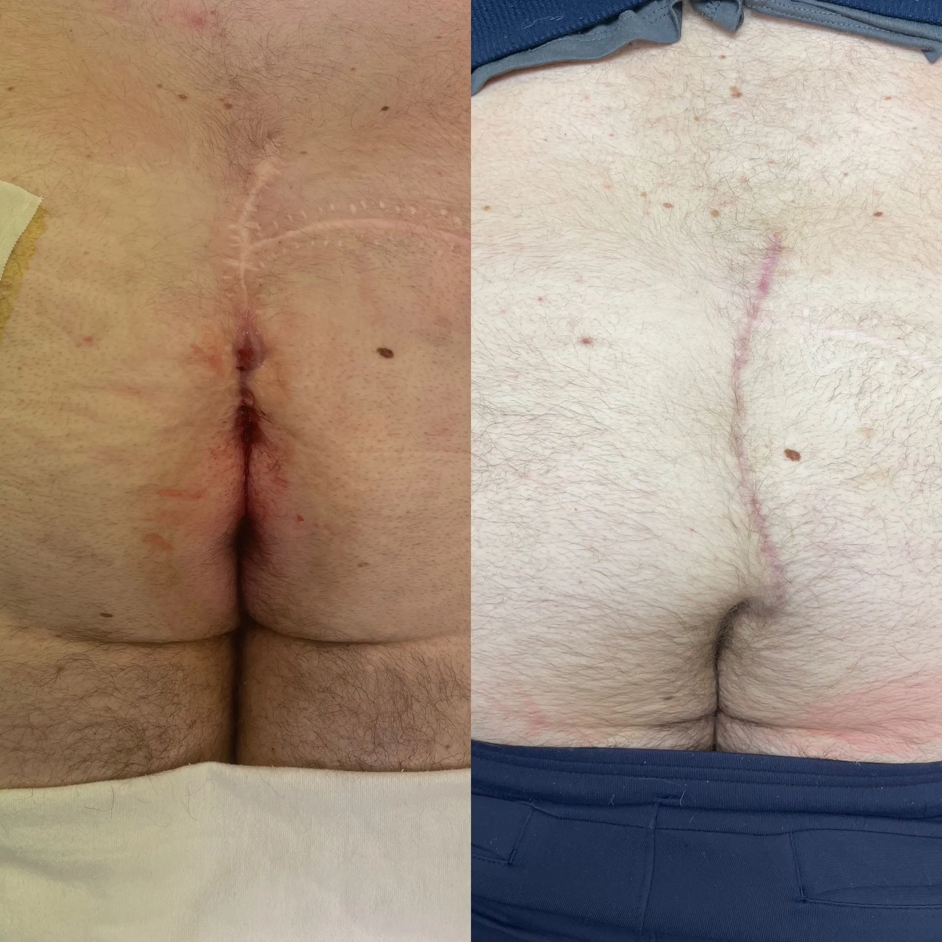
Patient 1
Before & After
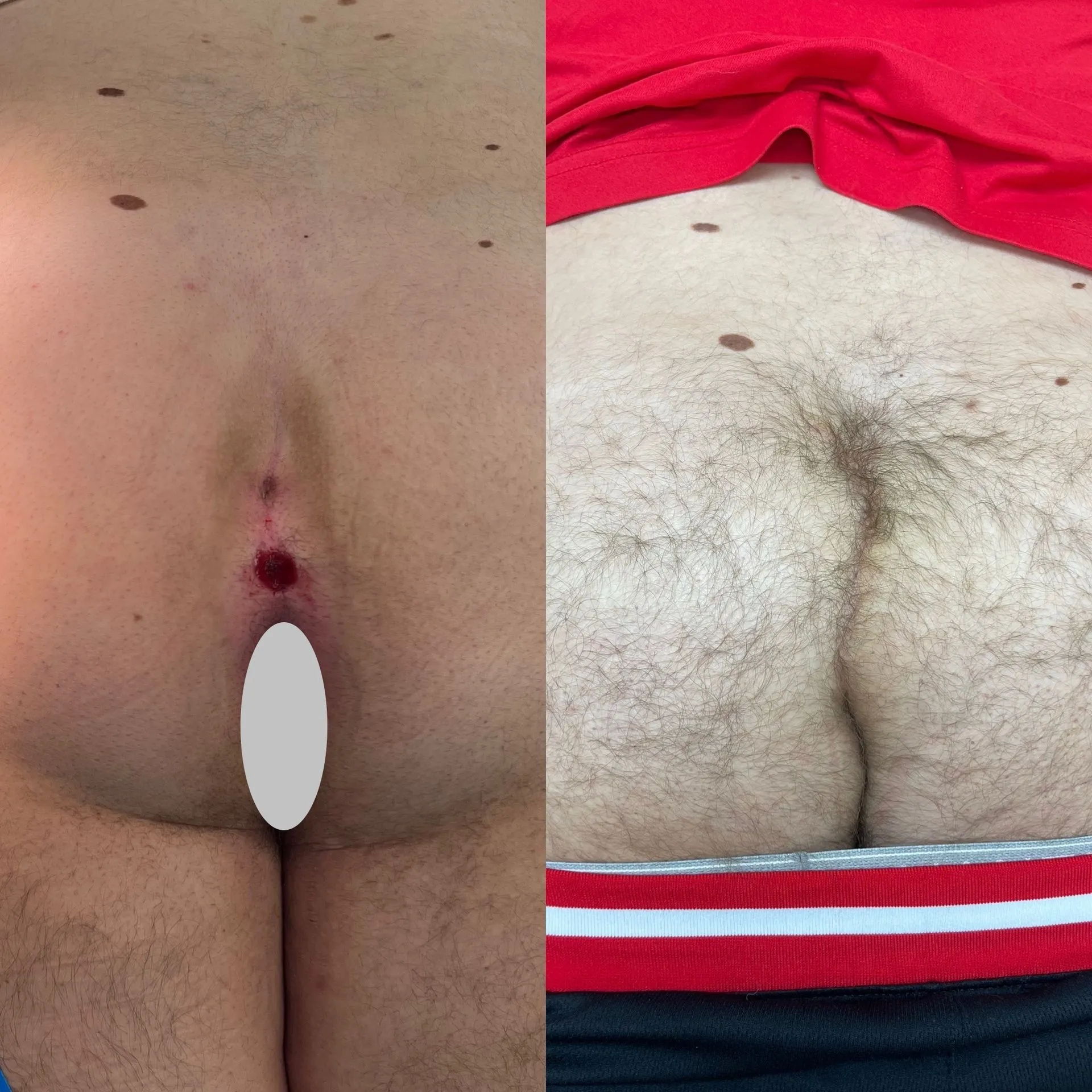
Patient 2
Before & After
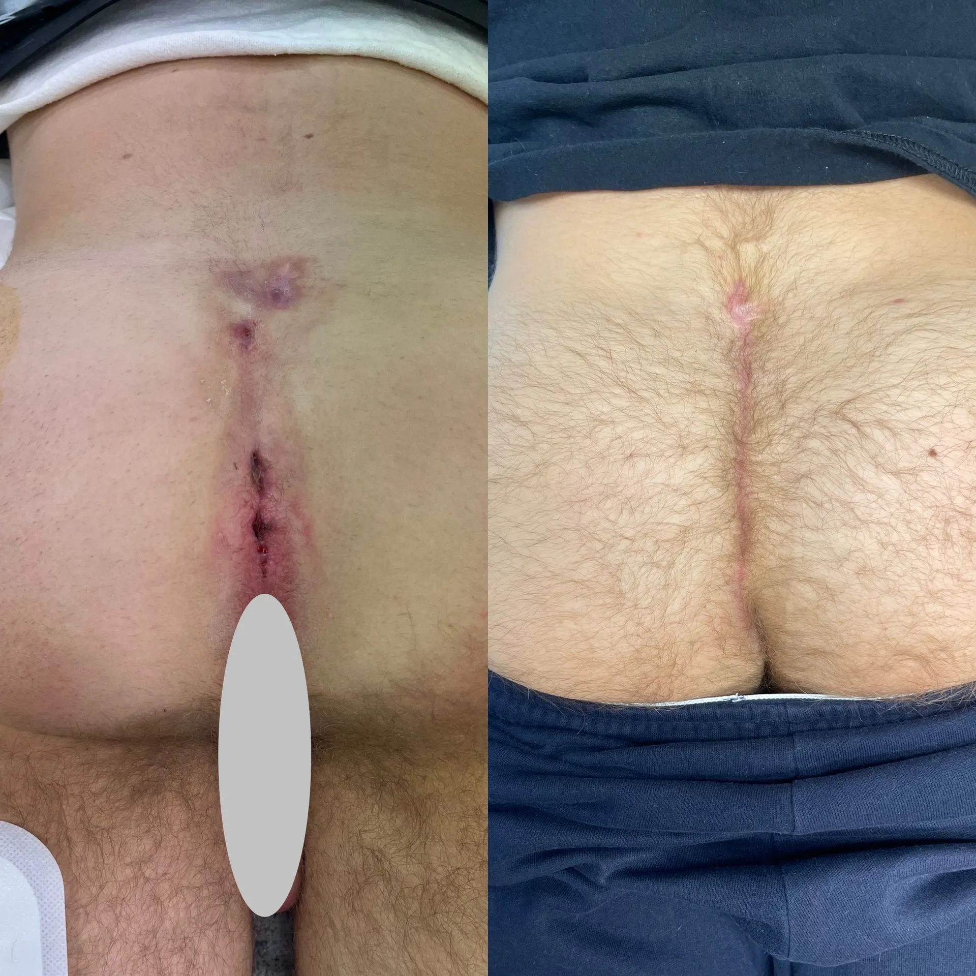
Patient 3
Before & After
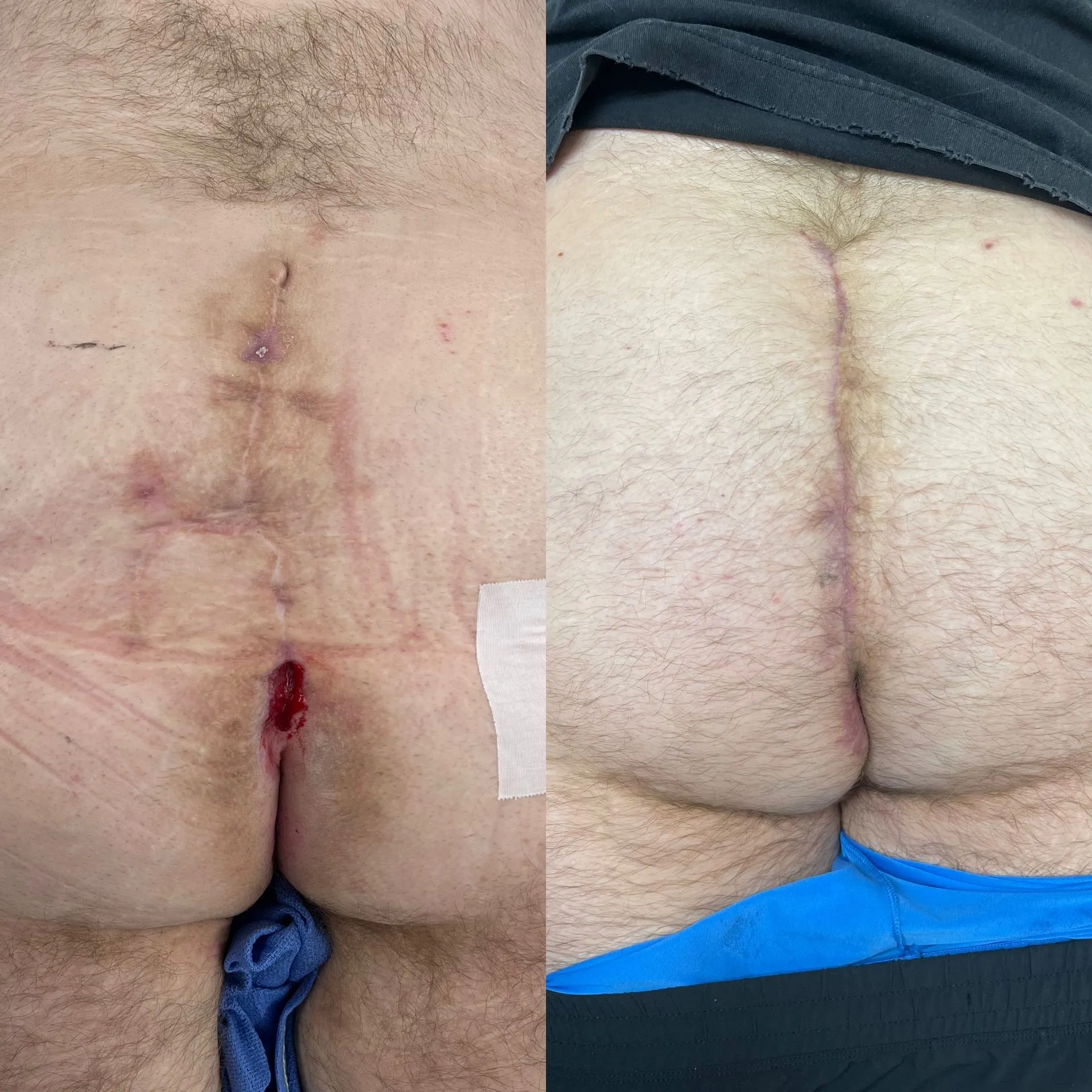
Patient 4
Before & After
Patient 5
Before & After
Patient 6
Before & After
Patient 7
Before & After
Patient 8
Before & After
Find Relief Today
Schedule Your
Consultation
Contact Pilonidal Fix today and let us help you leave your pain behind. Take the first step toward lasting relief with expert, compassionate care from Dr. Rafailov.
Call or Text
(973) 323-2400Hours
Mon–Thu 9–4, Fri 9:30–4